Reduction of periodontal pathogens adhesion by antagonistic strains
For figures, tables and references we refer the reader to the original paper.
The specific nutritional and physiological factors in the oral cavity give it a characteristic microflora that, most of the time, exists in a harmonious relationship with the host. This harmonious relationship can be disturbed, causing opportunistic pathogens to produce disease. One of the most common clinical manifestations of such a disturbance is periodontal disease. Periodontitis can develop when dental plaque accumulates at the gingival margin and the host reacts with an inflammatory response. Crevicular fluid flow is increased and as a consequence components of the immune system and different (glyco)proteins enter the crevice, from which the latter may act as a substrate for bacterial growth. Also, the crevice enlarges to become a pocket and the local environment becomes more and more anaerobic. The pocket fills not only with a mixture of microorganisms, but also with calculus, to which gram-negative obligate anaerobes may adhere (30). In time, infection and inflammation may spread from the gingiva to the ligaments and supporting bone to cause destruction of this connective tissue attachment of the teeth. Although periodontitis is the result of a multi-species biofilm, some species are considered as key periodontopathogens, including Porphyromonasgingivalis and Actinobacillusactinomycetemcomitans. Other species, for instance Prevotellaintermedia and Peptostreptococcus micros, are regarded as possible pathogens (1, 22, 31).
Therapeutic treatments are always aimed at removal of periodontopathogens from the subgingival area and worldwide-accepted strategies consist of scaling and root planning, sometimes supplemented with systemically or locally administered antibiotics. Yet, these treatments are often insufficient to cure a patient for prolonged periods of time, and hence alternative strategies to restore a healthy periodontal microflora are required (17). One such strategy could be the subgingival application of antagonistic strains that inhibit (re)colonization of the periodontal pockets by pathogenic bacteria. Early colonizers, like Streptococcus sanguinis and Streptococcus mitis, may possibly be regarded as antagonistic strains because of their in vivo/in vitro growth inhibition (7) and biosurfactant production (29), respectively. Alternatively, Streptococcus crista might be considered as antagonistic because it downregulatesFimA expression (32), while Streptococcus salivarius inhibits the emergence of mutans streptococci (26). Furthermore, the high prevalence of Haemophilusparainfluenzae and Actinomycesnaeslundii in periodontal health (10, 12) suggests that these may also be antagonistic strains.
The objective of this study was to identify bacterial strains that reduce the adhesion of periodontal pathogens to a surface. To this end, adhesion of periodontopathogens [P. gingivalis American Type Culture Collection (ATCC) 33277, P. intermedia ATCC 49046 and A. actinomycetemcomitans ATCC 43718] was studied in a parallel-plate flow chamber after preconditioning with the above mentioned, potentially antagonistic strains (S. sanguinis ATCC 49297, S. crista ATCC 49999, S. salivarius TOVE-R, S. mitis BMS, A. naeslundii ATCC 51655 or H. parainfluenzae ATCC 33966).
Material and methods
Microorganisms and growth conditions
Each strain was cultured anaerobically in 10 ml of its respective growth media (see Table 1) at 37°C in 80% N2, 10% H2 and 10% CO2, except S. mitis BMS which was grown aerobically. After 24 h, 200 ml medium was inoculated with the precultures and grown for 16 h under the appropriate conditions. Bacteria were harvested by centrifugation (6500 g, 5 min, 10°C), washed twice with working buffer (10 mm sodium phosphate and 154 mm sodium chloride, pH 7.0) and resuspended in working buffer, except P. gingivalis ATCC 33277 and P. intermedia ATCC 49046. These two strains were washed and resuspended in 40 mm potassium phosphate buffer, pH 6.9, in which no aggregation of bacterial strains or pairs of strains occurs during flow experiments. The suspensions were sonicated intermittently on ice for 30 s to break aggregates and chains, except for A. naeslundii ATCC 51655. Periodontopathogens were resuspended to a concentration of 3 × 10⁸ bacteria/ml and the antagonistic strains were resuspended to concentrations of 5 × 10⁷, 3 × 10⁸, and 1 × 10⁹ bacteria/ml.
Table 1.Bacterial strains and their growth media, as used in this study
Adhesion experiments
The parallel plate flow chamber (dimensions: l × w × h = 7.6 × 3.8 × 0.06 cm) and the image analysis system have been described in detail elsewhere (29). Briefly, the flow chamber consisted of a glass top and a polymethylmethacrylate (PMMA) bottom plate separated by two spacers with a thickness of 0.06 cm. Both plates were cleaned by sonication in 2% (volume/volume) RBS 35 detergent (Societé des TraitementsChimigues de Surface, Lambersat, France), rinsed thoroughly with tap water, dipped in methanol and rinsed again with tap water and demineralized water. For the manual enumeration of bacteria adhering to the bottom plate, bacteria were observed with a charge-coupled device camera (CCD-MXR, High Technology, Eindhoven, the Netherlands) mounted on a phase-contrast microscope equipped with a 40× ultra-long working distance objective, after image enhancement.
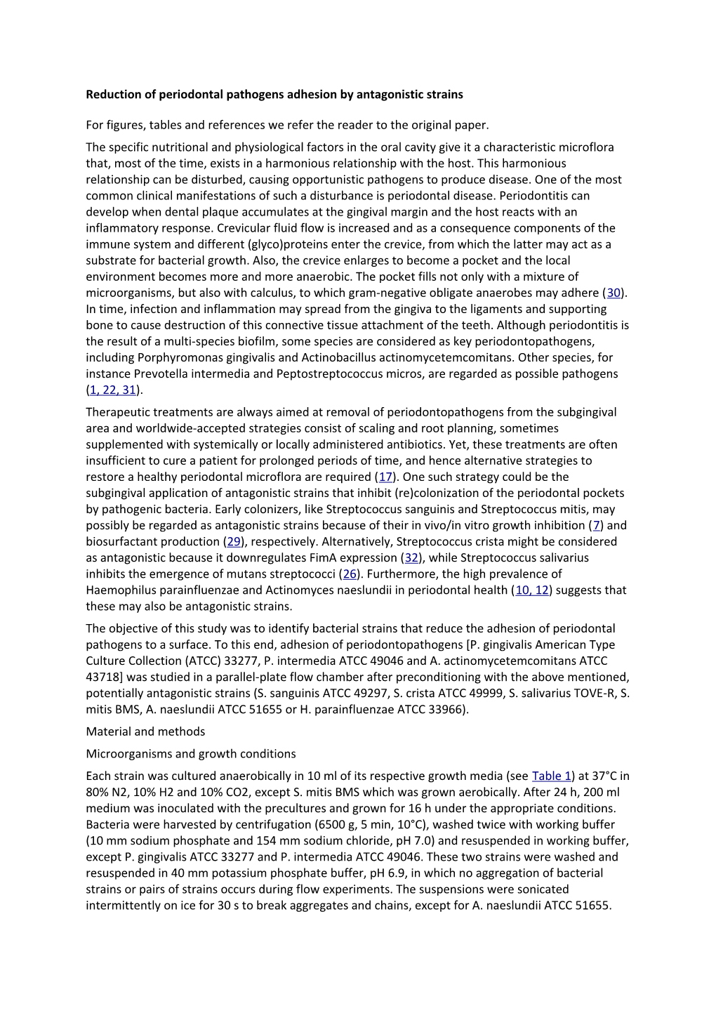
To study multiple antagonistic strains against a periodontopathogen in one experimental run, the so-called ‘dot assay’ was employed (11). In the ‘dot assay’, the PMMA bottom plate was pretreated in six discrete locations with the antagonistic strains. To this end, six 25-μl suspension droplets, i.e. two strains, each at three concentrations, were put on the PMMA bottom plate, as shown in Fig. 1. The closed chamber was left under slight agitation at room temperature for 30 min to allow sedimentation and adhesion of the bacteria. This procedure did not trigger the droplets to spread and consequently adhesion occurred only at the surface under the droplets. Next, the flow chamber was mounted on the stage of a phase-contrast microscope and connected to the flasks containing a periodontopathogen suspension and the appropriate buffer. All tubes and the flow chamber were filled with buffer, while care was taken to remove air bubbles from the system. The flasks containing buffer and bacterial suspension were positioned at the same height with respect to the chamber, so that immediately after the flows were switched, all fluids would circulate through the chamber under the influence of hydrostatic pressure at the desired shear rate of 10/s (0.025 ml/s), which yields laminar flow (Reynolds number 0.6).
Figure 1.
Schematic presentation of the unmounted parallel-plate flow chamber showing the positioning of the six 25-μl antagonistic suspension droplets on the bottom plate.
To remove the non-adhering antagonistic strains from the flow chamber, buffer was allowed to flow for 10 min through the flow chamber. It could be microscopically ascertained that the preconditioning procedure yielded bacterial adhesion only at the surface area of the bottom plate covered by the antagonistic suspension droplets (see Fig. 2A,B). Subsequently, images were taken to determine the number of pre-adhered antagonistic bacteria per cm2 on the bottom plate (Na) (see Fig. 2B). Thereafter, flow was switched for 2 h to a periodontopathogen suspension. Again, images were taken to determine the total number of bacterial cells per cm2, i.e. periodontopathogens and antagonist (Nt) in the case of focusing at the location previously covered by a suspension droplet (see Fig. 2D) or the number of periodontopathogens (Np) in the absence of antagonists (see Fig. 2C) when focusing on bare substratum surface, next to the previous location of the suspension droplets. It could be microscopically ascertained that no antagonist desorbed during the flow with periodontopathogens (note that all perfusion steps occurred under the same shear), therefore differential enumeration yielded the number of adhering periodontopathogens per cm2 in the presence of antagonistic cells (Np,a) by subtracting Na from Nt.
Figure 2.
Images taken within one experiment after pretreatment of a small part of the bottom plate of the parallel-plate flow chamber with a 25-μl suspension droplet of an antagonistic strain (upper series) and subsequently after 2 h of periodontopathogen flow (lower series). (A) Surface not pretreated with Streptococcus mitis BMS. Note that there are no adhering bacteria and conditioning is confined to the area underneath a suspension droplet (see B). (B) Adhering S. mitis BMS cells, i.e. antagonist surface coverage. (C) Adhering periodontopathogenActinobacillusactinomycetemcomitans ATCC 43718 in the absence of pre-adhering S. mitis BMS. (D) Adhering periodontopathogen A. actinomycetemcomitans ATCC 43718 in the presence of pre-adhering S. mitis BMS.
The decrease in adhering periodontopathogens, Rp, was defined as the reduction in the number of adhering periodontopathogen cells, achieved per adhering antagonistic bacterial cell; this could be expressed as
(1)
where Np is the number of adhering periodontopathogens per cm2 after 2 h in the absence of an antagonistic strain, Np,a is the number of adhering periodontopathogens per cm2 after 2 h in the presence of an antagonistic strain and Na is the number of pre-adhered antagonistic bacteria per cm2. By consequence, in the case of 106 adhering antagonists per cm2, an Rpof 2.5 cells/cell indicates that 2.5 × 10⁶ periodontopathogens have been prevented from adhering in comparison with bare substratum surface.
All adhesion experiments, both in the absence and presence of antagonistic strains, were performed at least three times with separately cultured organisms at room temperature. Deviations from zero in mean periodontopathogen reductions, Rp, were examined by Student’s t-test. The threshold level for significance was set at P < 0.05.
Results
In Table 2, the number of adhering periodontopathogens in the absence of antagonistic bacteria (Np) is presented for antagonist surface coverage less than 5%. Although antagonist surface coverage was achieved with different bacterial concentrations in suspension, the surface coverage was always less than 5% and, in addition, the mean reductions in periodontopathogen adhesion brought about per adhering antagonist were not significantly different. Hence in Table 2, no differentiation has been made with regard to antagonist concentration in suspension. As can be seen, these numbers vary between 5.2 × 10⁶, 4.3 × 10⁶, and 1.6 × 10⁶ cells/cm2 for P. gingivalis ATCC 33277, P. intermedia ATCC 49046, and A. actinomycetemcomitans ATCC 43718, respectively. The mean reductions in periodontopathogen adhesion per adhering antagonistic bacterium, Rp, are also shown in Table 2. As a result of experimental errors in enumeration, Rp values can become slightly negative (i.e. adhesion of P. intermedia ATCC 49046 in the presence of S. sanguinis ATCC 49297), but never significantly different from zero, in contrast to the large reduction achieved in adhesion of 3.8 P. gingivalis ATCC33277 cells by one adhering A. naeslundii cell.
Table 2.Number of adhering periodontopathogens (Np) and the effect of antagonistic strains on periodontopathogen adhesion (Rp) to the bottom plate of the parallel-plate flow chamber
Of the three periodontopathogens P. gingivalis ATCC 33277 was generally inhibited most, especially by A. naeslundii, H. parainfluenzae, S. mitis, and S. sanguinis, but S. crista hardly inhibited this strain. Adhesion of P. intermedia ATCC 49046 was less effectively inhibited than that of P. gingivalis, and S. mitis caused the highest significant reduction in adhesion of P. intermedia ATCC 49046. A. actinomycetemcomitans ATCC 43718 was most difficult to inhibit, and its highest significant Rp value was caused by S. salivarius.
Discussion
Adhesion to host surfaces is the first step in infection. The possibility that antagonistic organisms could be used to control the adhesion of pathogens and prevent infection is called ‘replacement therapy’. For example, insertion of the antagonistic, α-hemolytic streptococcal strain 215 in the pharynx of a group of infants lacking this α-hemolytic species, yielded a strong reduction in the number of the bacterial pneumonia cases in this group compared with a control group (24). Another example, although thus far only tested in vitro, is the construction of an effector strain of Streptococcus mutans for replacement therapy of dental caries. In vitro experiments showed a selective advantage in colonizing the teeth and little or no cariogenic potential (7).
In the literature it is pointed out that antagonistic strains are better adapted to their niche than potential pathogens, and can therefore interfere in disease by passively occupying the niche, actively restricting the adhesion capability of pathogens to surfaces (blocking), negatively influencing the vitality or growth of pathogens (more competitive for essential nutrients), and/or adversely modifying the production of or degrading the virulence factors of the pathogens (6, 23). However, definitive proof that any of these mechanisms occur has seldom been given. One well-documented example is the competition for essential nutrients, which explains, at least in part, the potential of the α-hemolytic streptococcal strain 215 to reduce colonization by Streptococcus pyogenes (19).
In this study the capacity of six antagonistic bacteria to block the attachment of periodontopathogens, i.e. reduce the adhesion of periodontopathogens, was evaluated. For quantification of blocking we chose a simple model, namely a comparison of the number of adhering periodontopathogens to a substratum surface per unit area in the absence and presence of an antagonistic strain. A good device for studying blocking of attachment in vitro is the parallel-plate flow chamber with in situ observation and image analysis options (29) because it offers the chance to calculate the physical parameters of the blocking process, such as the number of blocked pathogens per adhering antagonist cell or to account for shear-off forces, like for instance those as a result of salivary flow. Moreover air–liquid interface passages are avoided, yielding accurate enumeration of the number of adhering bacteria under flow conditions and the antagonistic strains can be easily introduced at the start of the experiment.
Taking stock of the results presented in Table 2 it appears that A. naeslundii, H. parainfluenzae, and S. mitis cause the strongest blocking of P. gingivalis adhesion but have considerably less effect on the adhesion of P. intermedia and A. actinomycetemcomitans, suggesting that the blocking effect is caused by interactions, specific to each combination of an antagonist and periodontopathogen. Indeed, blocking is determined by repulsive interactions between the adhering antagonist and the periodontopathogen (4, 21) and larger blocking effects may arise, for instance, from biosurfactant production. For S. mitis BMS blocking of attachment by biosurfactant production has been reported (29), which might be unique to S. mitis among the current collection of antagonistic strains and could be more effective than the interference mechanisms described hitherto for S. sanguinis, S. crista, and S. salivarius (7, 27, 32). Interestingly, previous reports showed, instead of antagonistic effects, synergistic effects of A. naeslundii on P. gingivalis through co-aggregation (33) and enhancement of P. gingivalis adhesion (20). However, because it is clinically known that Actinomyces occurs more in healthy pockets than in diseased pockets (12), we believe that the A. naeslundii strain studied here must be considered as an antagonist of P. gingivalis.
Periodontitis is an infectious disease and is of major concern for both dentists and patients, affecting at least one-third of the population worldwide (13, 15). Treatment is focused on removal of the periodontopathogens and the subsequent, long-term development and maintenance of a non-pathogenic microflora in the subgingival area by scaling and root-planning, sometimes supplemented with antibiotics and a conscientious program of oral hygiene by tooth brushing and interdental cleaning via toothpicks and/or brushes. Unfortunately, many patients remain at risk for periodontitis partly because of their lack of compliance, yielding recolonization of the pockets by pathogenic bacteria that survived the therapy and/or became dislocated from other niches, such as untreated sites, tongue, and saliva (16). In medical fields other than periodontology, research efforts into the clinical effects of antagonistic strains in man are increasing and a number of health benefits have been postulated (see Table 3 for a summary).
Table 3.Postulated beneficial effects of intake/installation antagonistic strains in man
In the present study, we show that adhesion of selected periodontopathogens can be inhibited by antagonistic strains, most notably by S. mitis BMS. This strain would thus be a good candidate for ‘replacement therapy’.
Acknowledgment
This study was supported by the NIH, grant number 1R21 DE015360-01.